
[NS1] Neurons and Glia
안녕!
이번 시간에는 신경과학의 기초 중의 기초!
바로 신경세포(neuron)과 신경아교세포(glial cells)에 대해 다룰 예정이다!
The Neuron Doctrine
Finding Neuron via Histology
Histology: 염색법
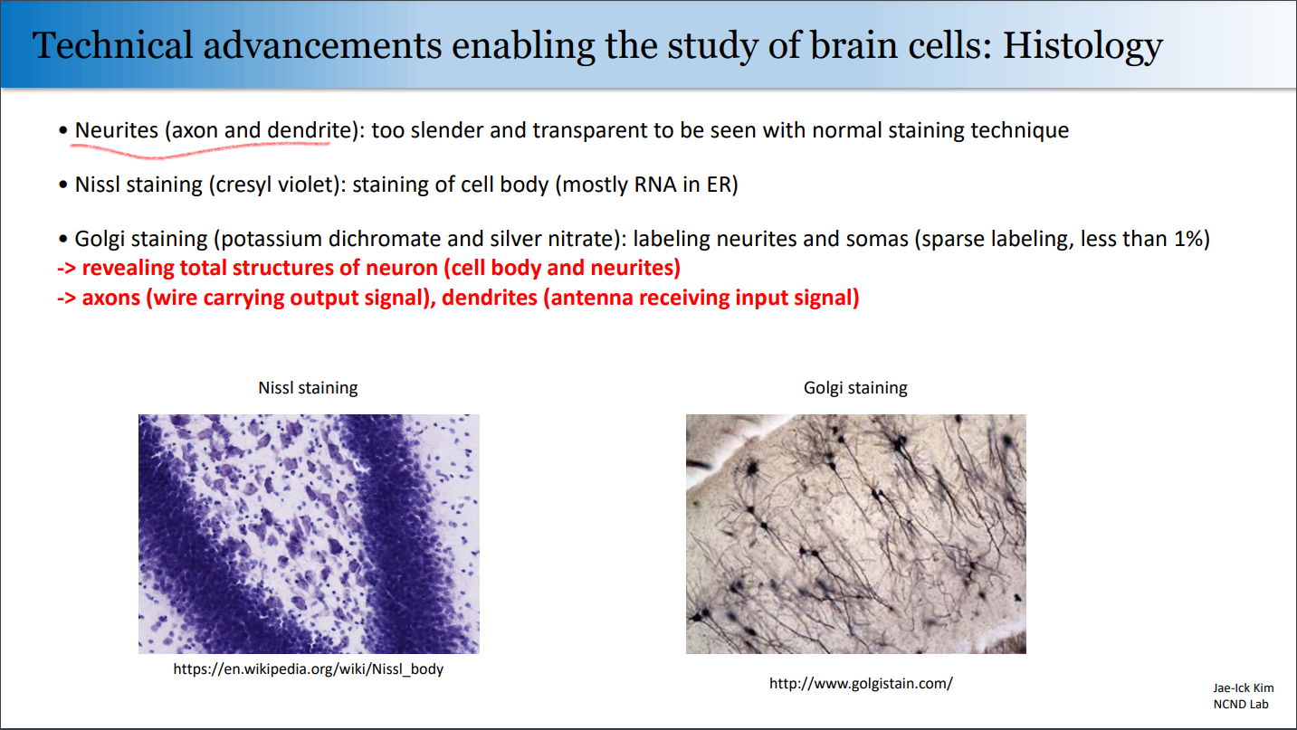
그래서 대충 2가지 염색법이 있는데,
바로 Nissl Stain과 Golgi Stain이다.
Nissl Stain
❗단어들❗
| 단어 | 뜻 |
|---|---|
| stain | 얼룩, 염색 |
| clump | 1. 수풀/나무숲 2. (사람/사물)집단, (흙)덩어리 |
A German neurologist Franz Nissl showed that
a class of basic dyes would stain the nuclei of all cells as well as clumps of material surrounding the nuclei of neurons.
Nissl Bodies: The clumps stained by the dyes.
(cf. mostly RNA in ER)
Nissl Stain: The stain.
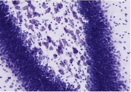
Useful for…
- Distinguish between neurons and glia
- Enables histologists to study the arrangement (cytoarchitecture) of neurons in different parts of the brain.
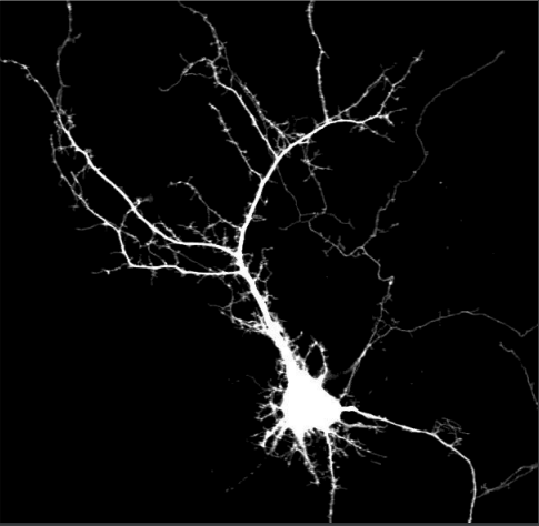
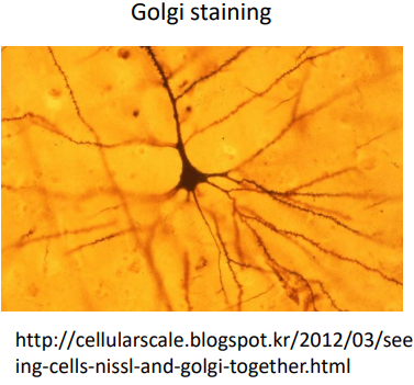
Golgi Stain
A silver chromate solution darkly colors the brain tissue.
It reveals the total structure of neuron, as follows…
Apparent Structure of Neuron
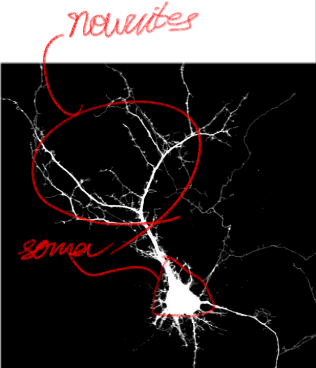
- 몸뚱아리: Soma (plural: somata) == Cell Body == Perikaryon (plural: perikarya)
- 수염: Neurites. (2 kinds)
Neurite 종류
- axons: [Output]
- Usually one for each cell body
- Of uniform diameter throughout its length
- Branch extensions are generally at right angles
- Can be long. >=1m
- dendrites: [Input]
- Extends from the cell body.
- Generally taper to the fine point.
- Comes in contact with many axons.
- Rarely longer than 2mm.
.png)