
[NS18] Brain Rhythms and Sleep
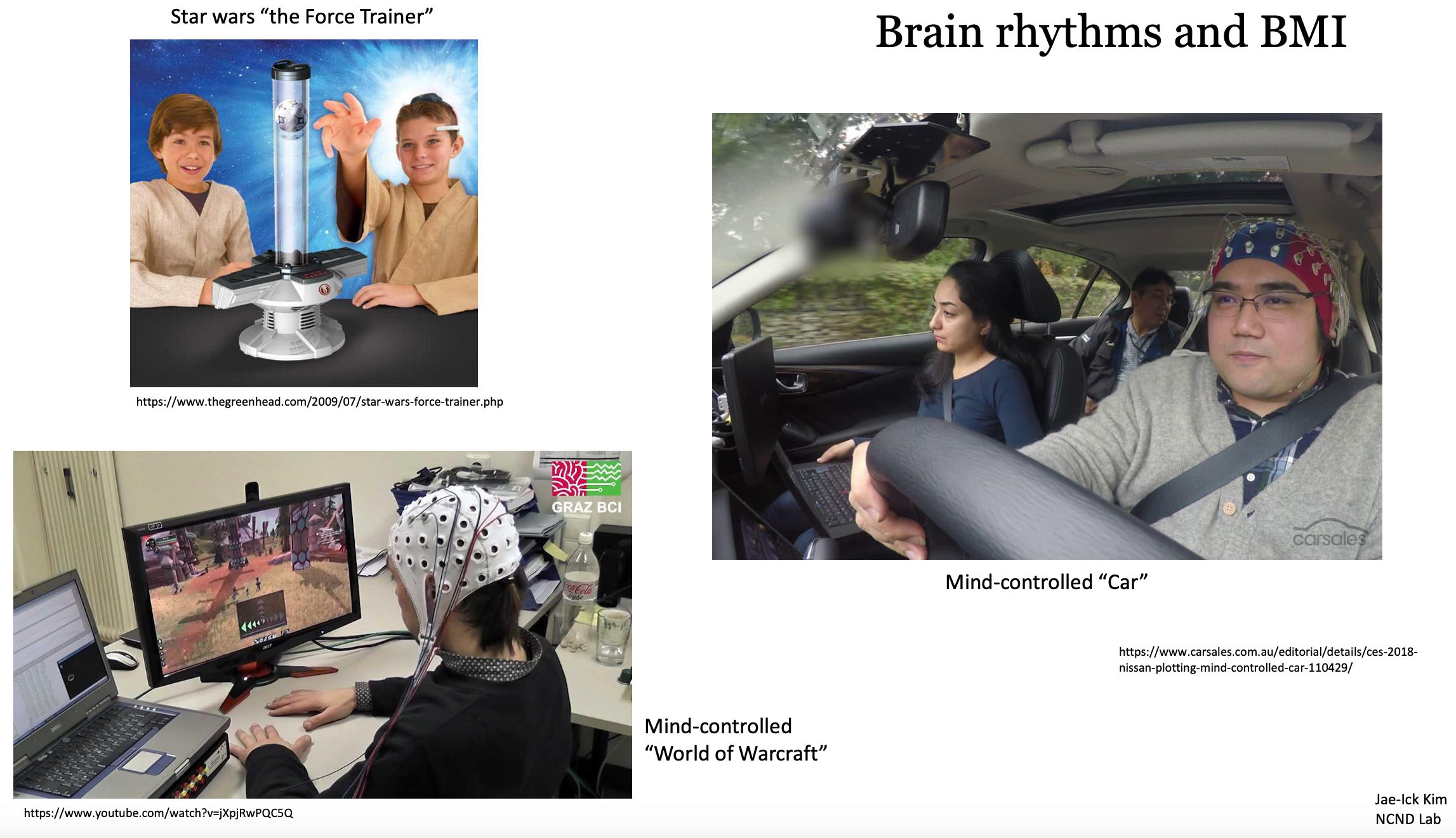
Brain Rhythms and BMI: Brain-Machine Interface
What we discusss:
- Explore selected brain rhythms, beginning from the fast to the slow ones.
- Discuss the elecroencephalogram (EEG); classical method of recording brain rhythms + sleep
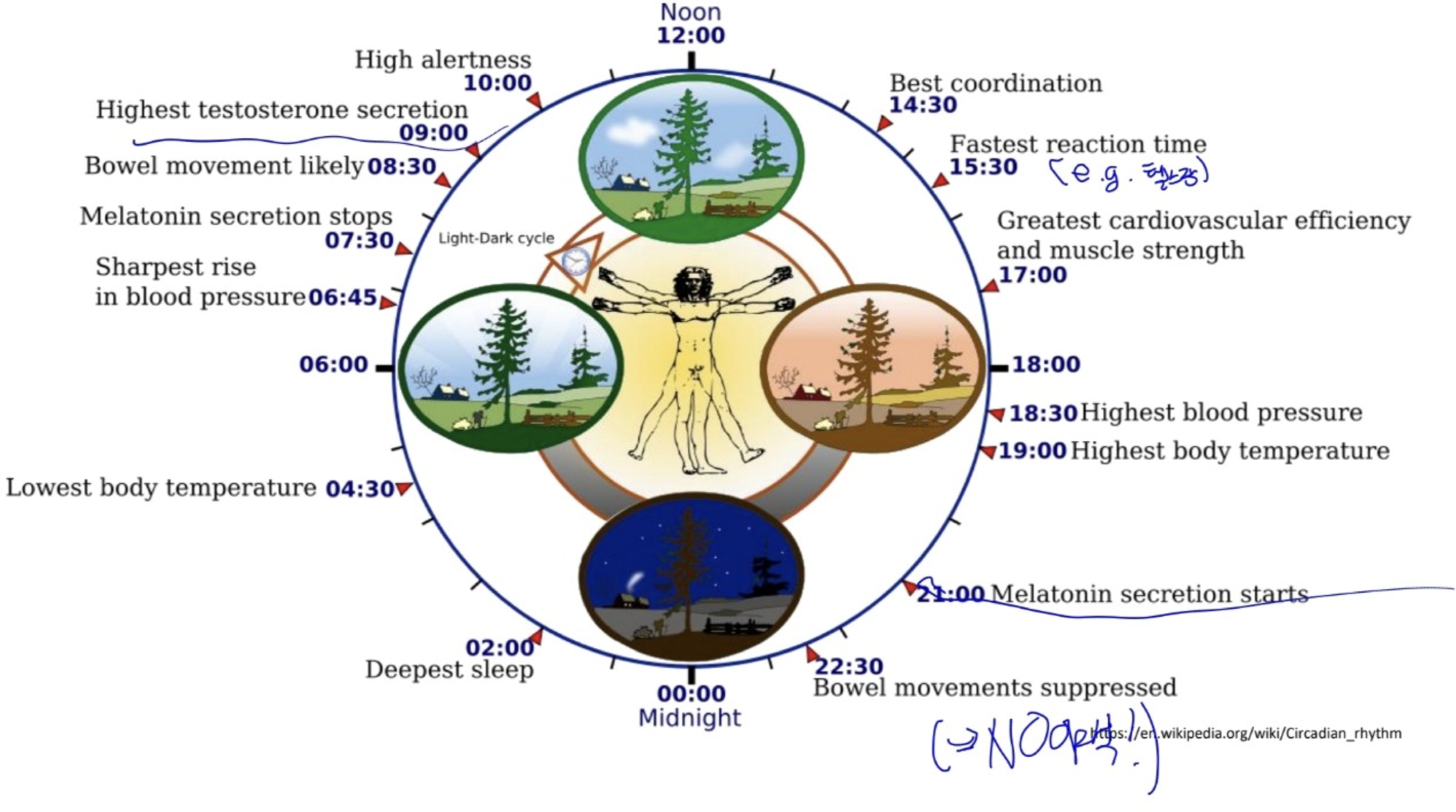
- Summarize circadian rhythms
1. The Electroencephalogram
Terms:
-
- Electroencephalogram(EEG)
- Measurement of electrical activity from the surface of the scalp that enables us to glimpse the generalized activity of the cerebral cortex.
- First described by Hans Berger (1929) (sleeping and waking EEGs are distinctly different)
- introduced by English physiologist Richard Caton in 1875. </span>
- Usage: study…
- help diagnose certain neurological conditions (seizures of epilepsy)
- study sleep and cognitive processes during wakefulness
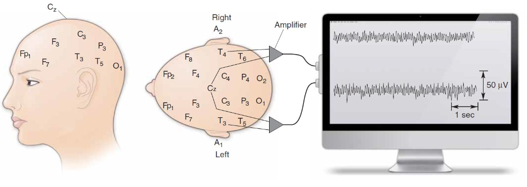
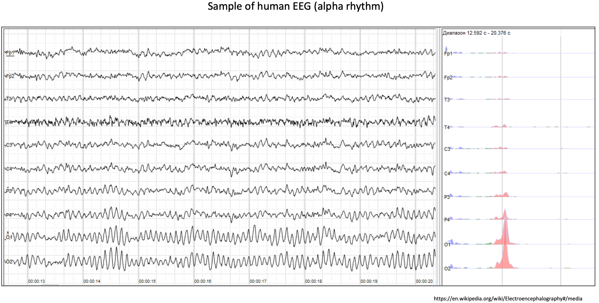
1.1. Recording Brain Waves
How to record? Simple:
- wear EEG electrodes, wires taped to the scalp along with conductive paste for a low-resistance connection.
- Pick two electrodes and measure the voltage difference => a few tens of microvolts.
What generates the fluctuations and oscillations of an EEG? (중요!)
문장 그대로! (in test)
The amplitude of the EEG signal strongly depends on the synchronousity of the activity of the underlying neurons. (and the number of neurons, of course)
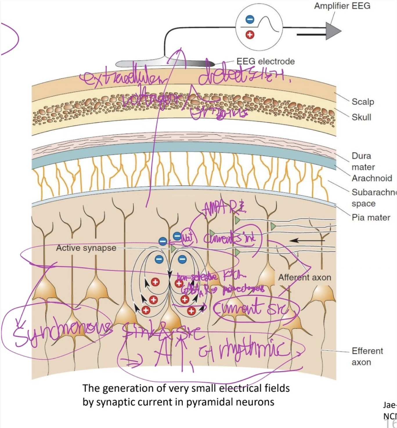
Generation of very small electrical fields by synaptic currents in pyramidal cells
When the afferent axon fires, the presynaptic terminal releases glutamate, which opens the AMPA receptor, a cation (Na+) channel.
Positive current flows into the dendrite, leaving a slight negativity in the extracellular fluid.
(by some non-selective K+ channel and GABA_A_R) the current escapes from the deeper part of the dendrite, leaving the fluid slightly positive.
The EEG electrode measures this pattern through thick tissue layers.
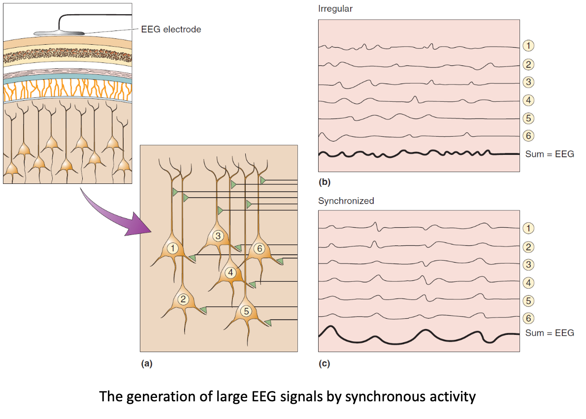
The generation of large EEG signals by synchronous activity.
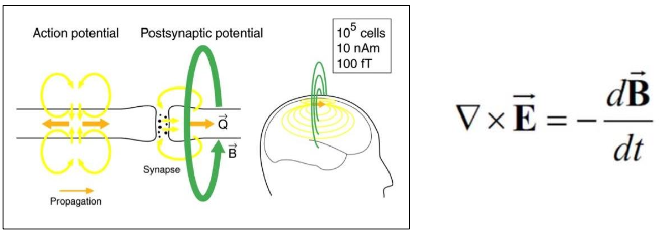
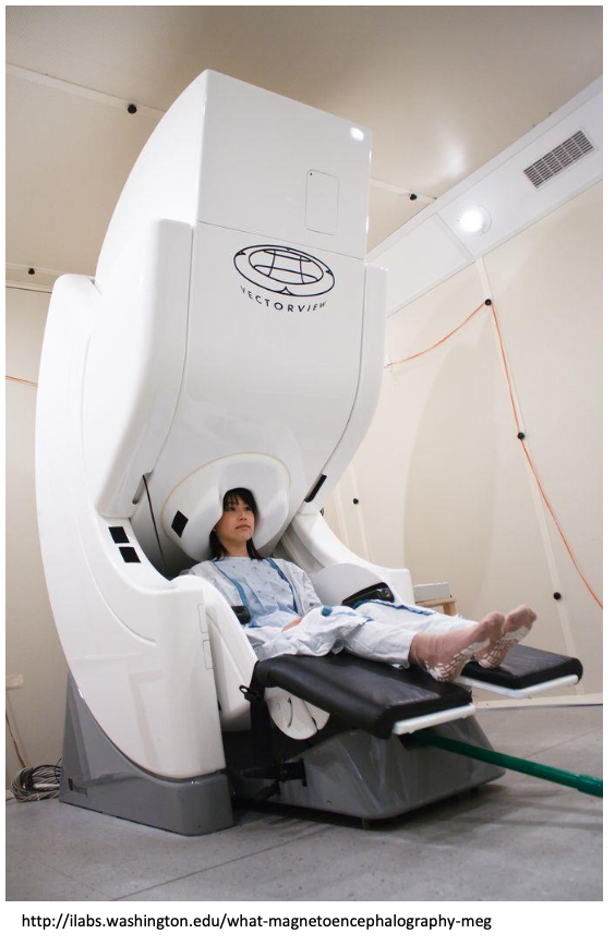
magnetoencephalography(MEG)
- Alternative way to record brain rhythms of cerebral cortex.
- Neurons generate currents => produce a magnetic field. (Faraday’s law of induction, or Faraday-Maxwell equation)
- Very small amplitude
- Requiring shielding room and highly sensitive magnetic detectors cooled at -269°C (with He(l) ).
Some features of MEG
1) Better than EEG at localizing the sources of neural activity in the brain 2) Can record rapid fluctuations of neural activity (like EEG, unlike fMRI or PET)
Differences with fMRI or PETs
1) No spatially detailed images of fMRI. 2) EEG and MEG directly measure the activity of neurons. (unlike fMRI and PET that detect changes in blod flow or metabolism, which may be influenced by other psysiological factors other than neuronal activities.)
cf. Electrophysiology: recording neuronal activities
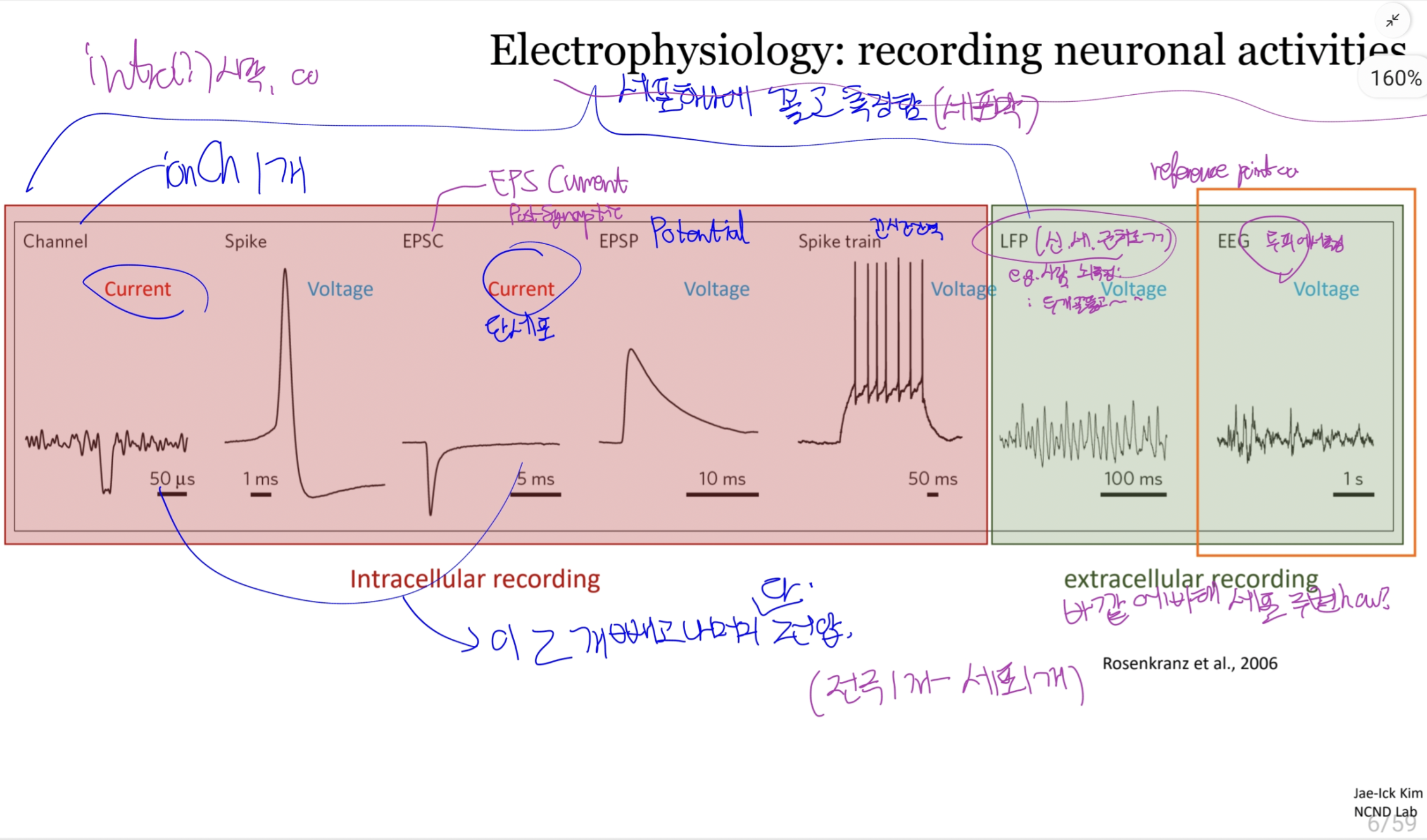
(dura, arachnoid, pia) ECoG(Electrocorticography)는 두개골 뚫고 경막(dura mater) 밖 (or 안)에서 Voltage를 측정하는 그런 것이다.
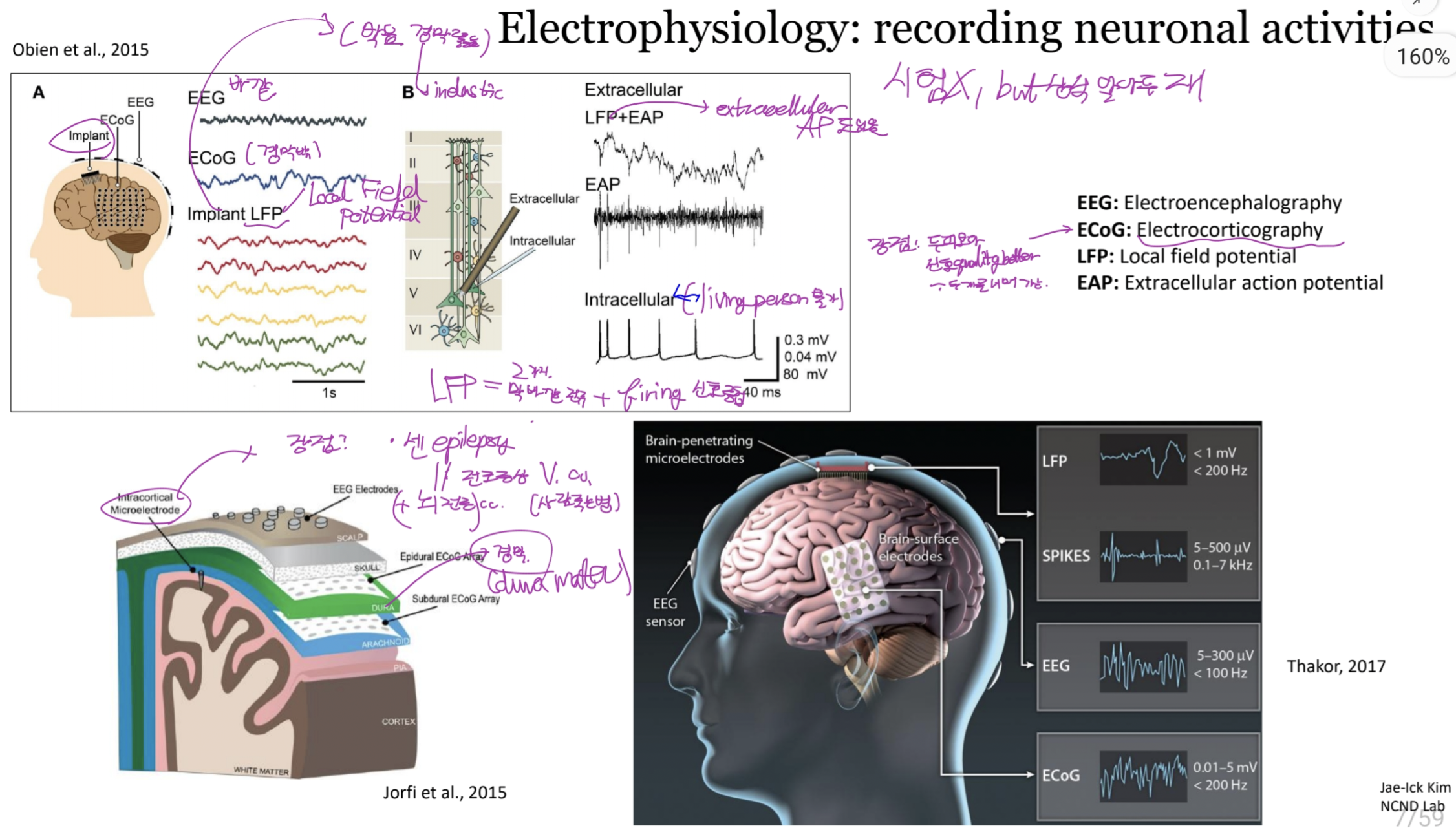
- Epidural ECoG Array: 경막 밖 표면
- Subdural ECoG Array: Arachnoid membrane 밖 표면
등등
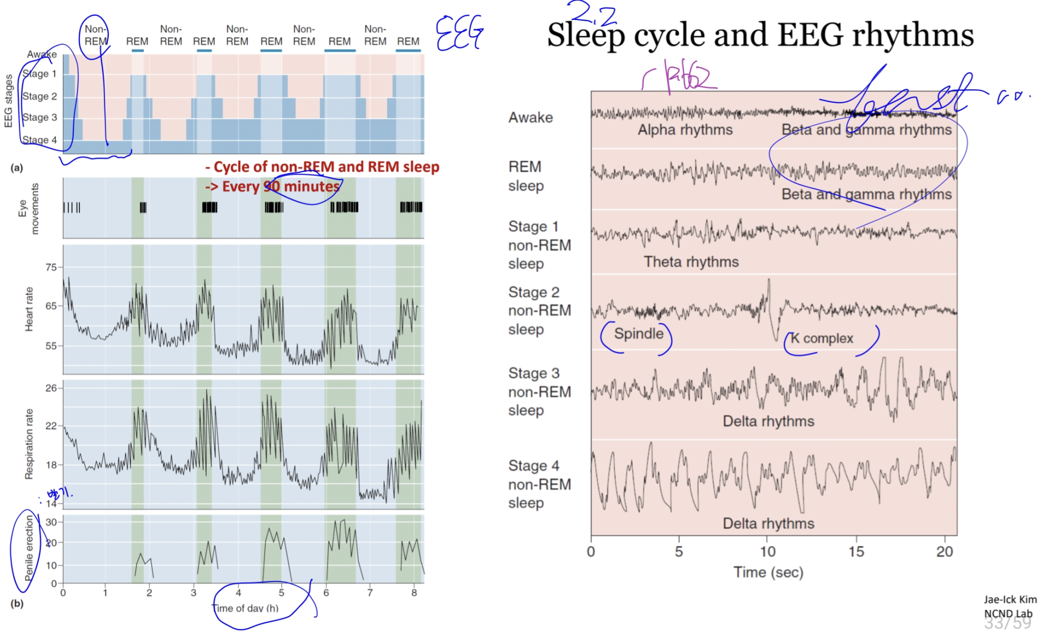
1.2. EEG Rhythms
EEG rhythms
- Correlated with brain states (attentiveness, sleeping, waking) and pathological conditions (seizures or coma)
- Normal EEG: very slow (0.05Hz)-very fast (>500 Hz)
- Additional rhythms: spindles (brief 8-14 Hz waves associated with sleep), ripples (brief bout of 80-200Hz oscillations)
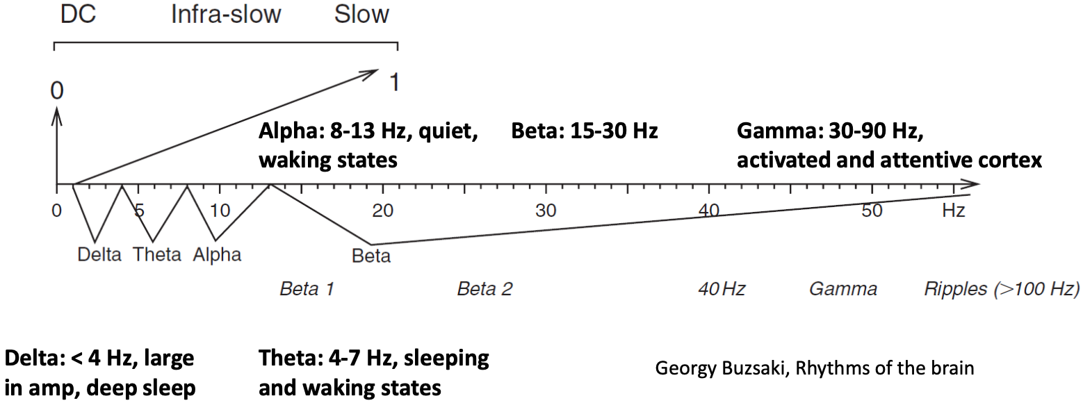
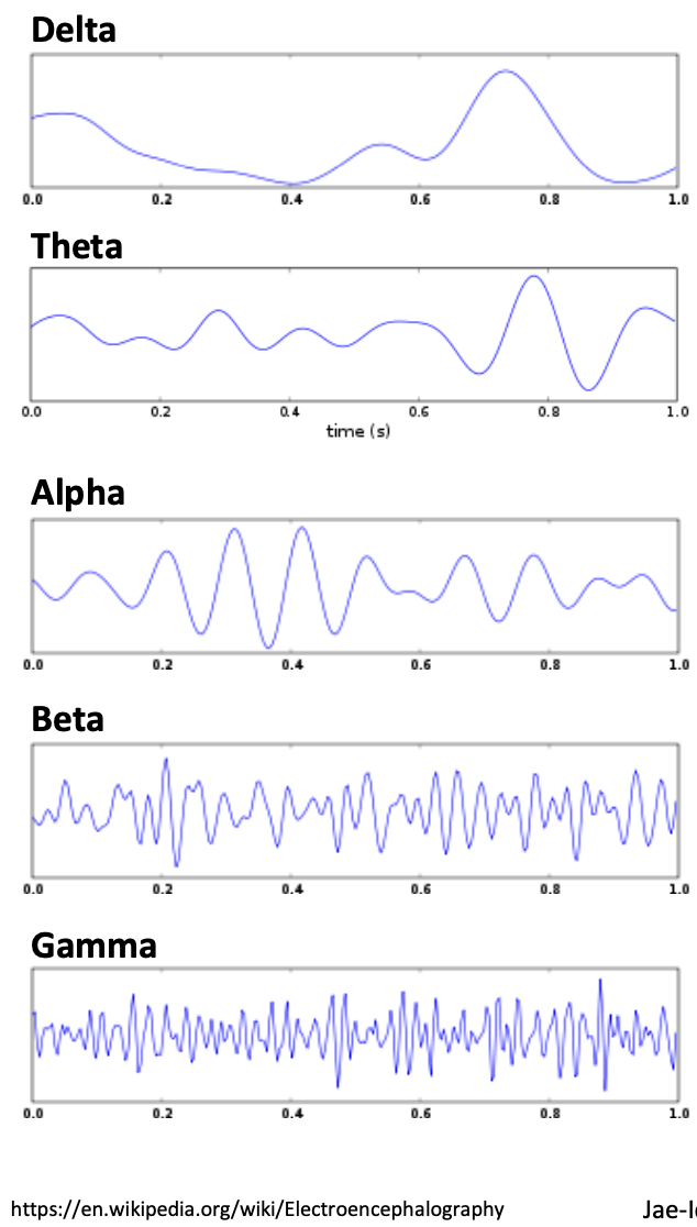
Frequencies
| δ | θ | α (mu) | β | γ |
|---|---|---|---|---|
| 0-4 | 4-7 | 8-13 | 15-30 | 30-90 |
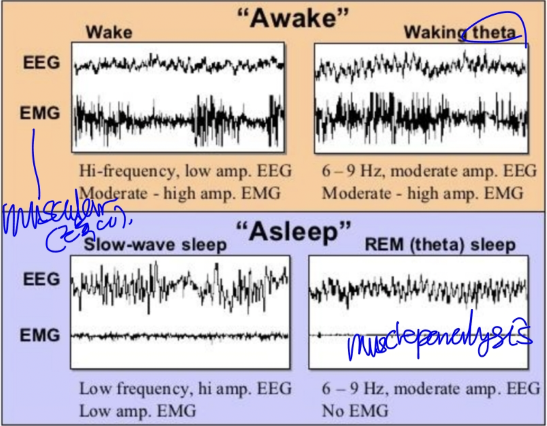
In general,
- High-frequency, low-amplitude rhythms are associated with alertness and waking, or the dreaming stages of sleep.
- Low-frequency, high-amplitude rhythms are associated with nondreaming sleep states, certain drugged states, or the pathological condition of coma.
Reasons? Later. (p.653)
1.3. Mechanisms and Meanings of Brain Rhythms
1.3.1. The Generation of Synchronous Rhythms
Two mechanisms of synchronous rhythms:
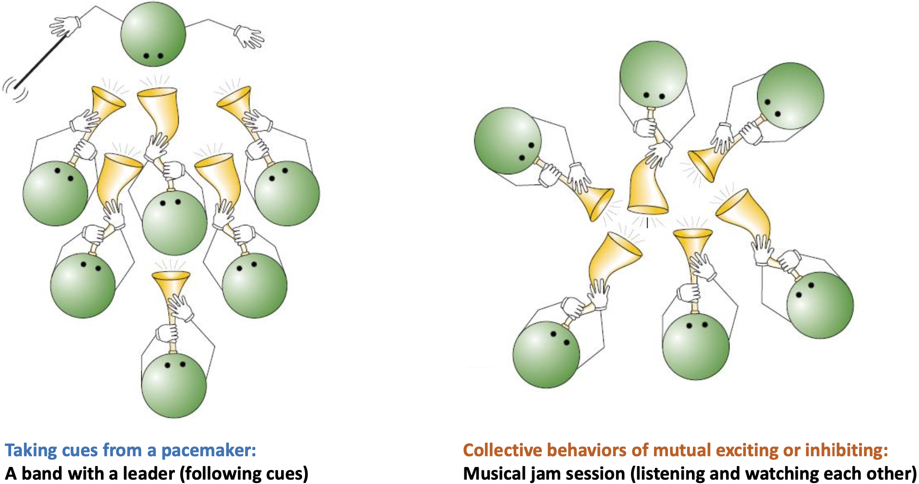
- Leading by a pacemaker (following cues)
- Collective behaviors of mutual exciting or inhibiting (listening and watching each other)
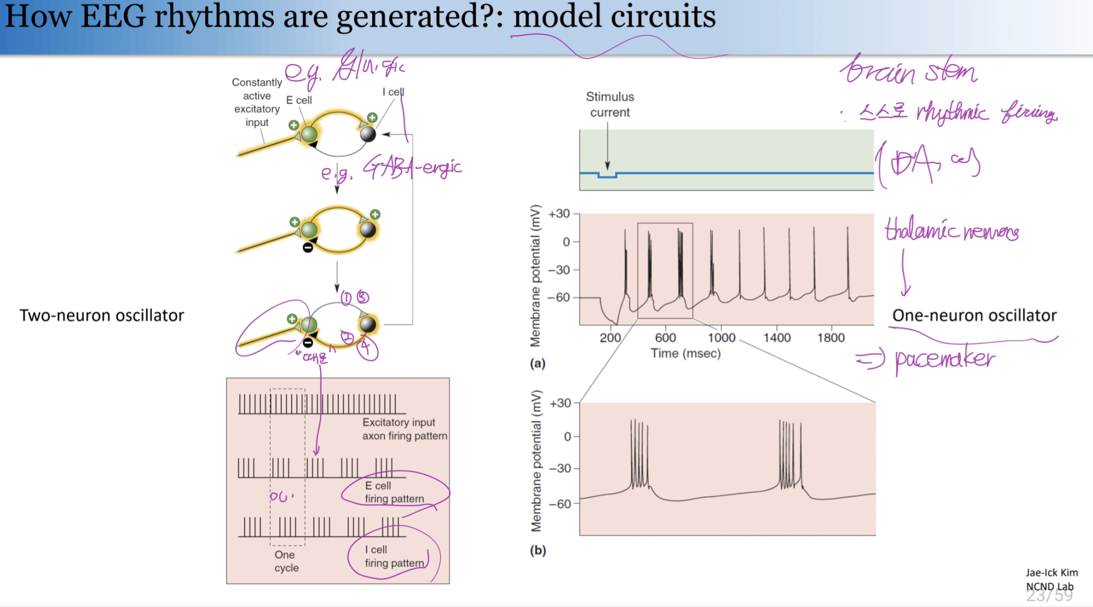
How EEG rhythms are generated?: model circuits
Two-neuron oscillator and one-neuron oscillator. (thalamic neurons can fire in patterns that do not reflect their input)
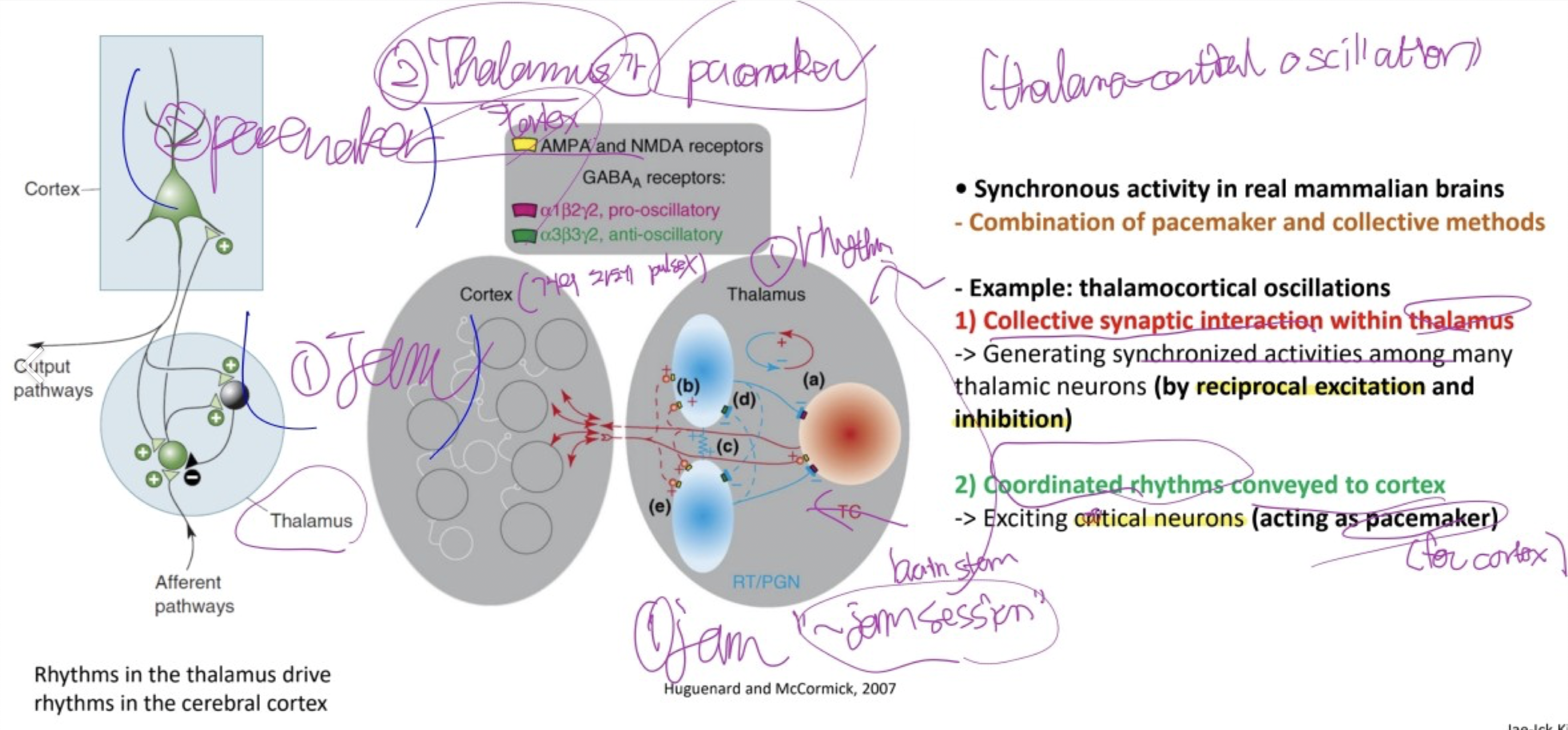
The synchronous activity in real mammalian brains is exhibited by the combination of pacemaker and collective methods:
e.g. Thalamocortical oscillations
1) The collective synaptic interaction within thalamic neurons generate synchronized activities among many thalamic neurons (by reciprocal excitation and inhibition) 2) The coordinated rhythms are conveyed to cortex, acting as a pacemaker, to excite the cortical neurons.
1.3.2. Functions of brain rhythms
Unknown… three hypotheses:
- Way to disconnect the cortex from sensory input.
- e.g. sleep-related, self-generated rhythms prevents sensory information from being relayed to the cortex.
- Drawback: Necessity of rhythms. (e.g. why not just inhibit the thalamus and let the cortex rest quitely?)
- Way to coordinate activity between regions of nervous system
- e.g. Fast rhythms in the awake cortex.
- Synchronously activity of cortical neurons responding to the same object in visual perception.
- Both sensory and the motor systems of the awake brain generate bursts of synchronous neural activity that give rise to EEG gamma rhythms (30-90Hz)
- Synchronizing fast oscillations bind together various neuronal components into a single perceptual construction. e.g. encoding “basketballness”
- Drawback:
- Indirect evidence, not proven, controversial.
- e.g. Fast rhythms in the awake cortex.
- By-products of highly-interconnected brain circuits. i.e. no function.
- Unavoidable consequence of varioous forms of feedback circuits.
- EEG rhythms can still provide us with a convenient windowon the functional state of the brain.
1.4. The Seizures of Epilepsy
Seizures: The most extreme form of synchronous brain activity. Firing with abnormal synchrony (and very large EEG patterns)
This is always a sign of pathology.
cf. Generalized and partial seizures
- generalized seizure: involving entire cerebral cortex of both hemispheres,
- partial seizure: involves only a circumscribed area of the cortex.
Epilepsy: The condition that a person experiencing multiple seizures (“isolated” seizures neglected.). (~= symptom of a disease.)
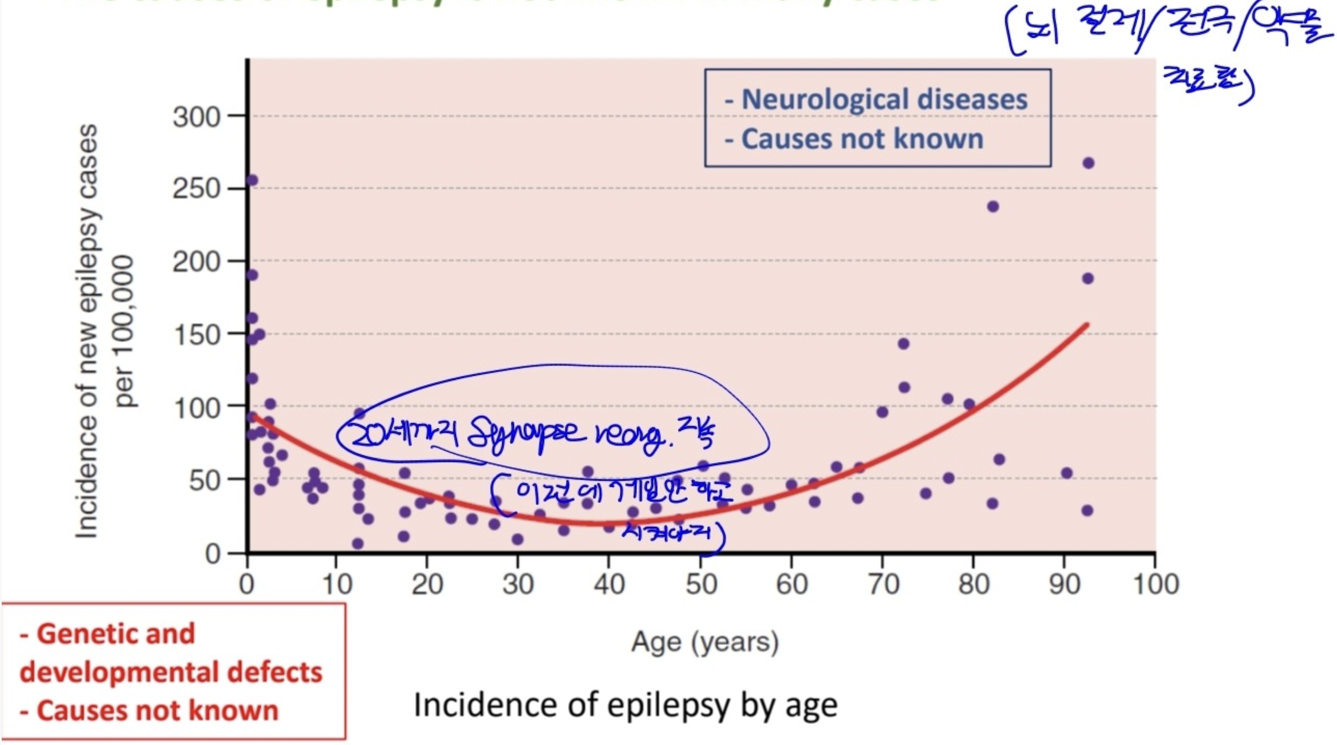
- Diagnozed most often in young children and the elderly.
- Causes: unknown in many cases.
- cogenital: genetic factors exists.
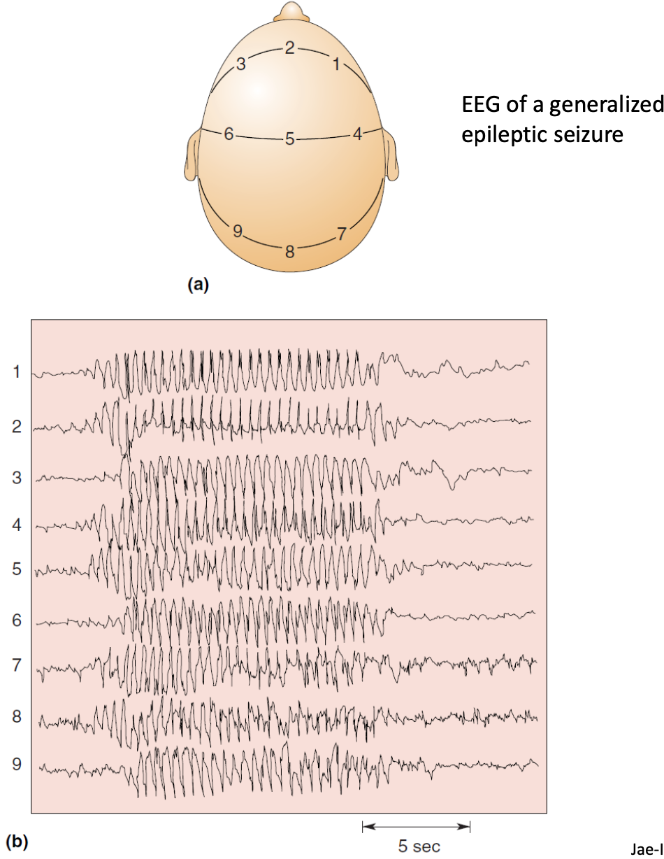
EEG of a generalized epileptic seizure
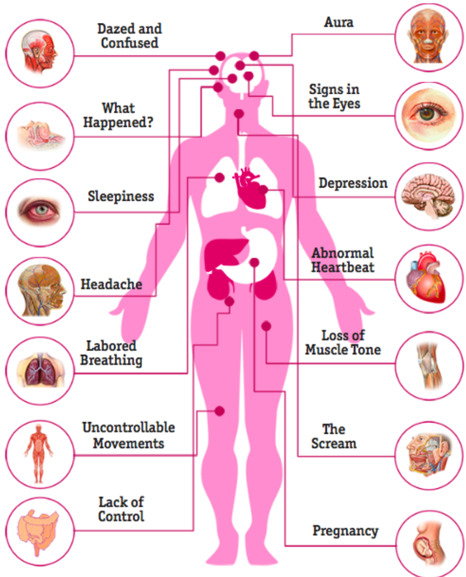
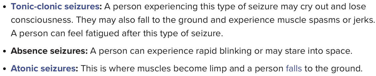
cf. Generalized onset seizures
retrieved from https://www.medicalnewstoday.com/articles/seizure-orgasm#overview
Mechanisms of Epilepsy
Molecular and cellular mechanisms of epilepsy
- Genetic mutation and predisposition: ion channels, transporters, receptors, signaling molecules
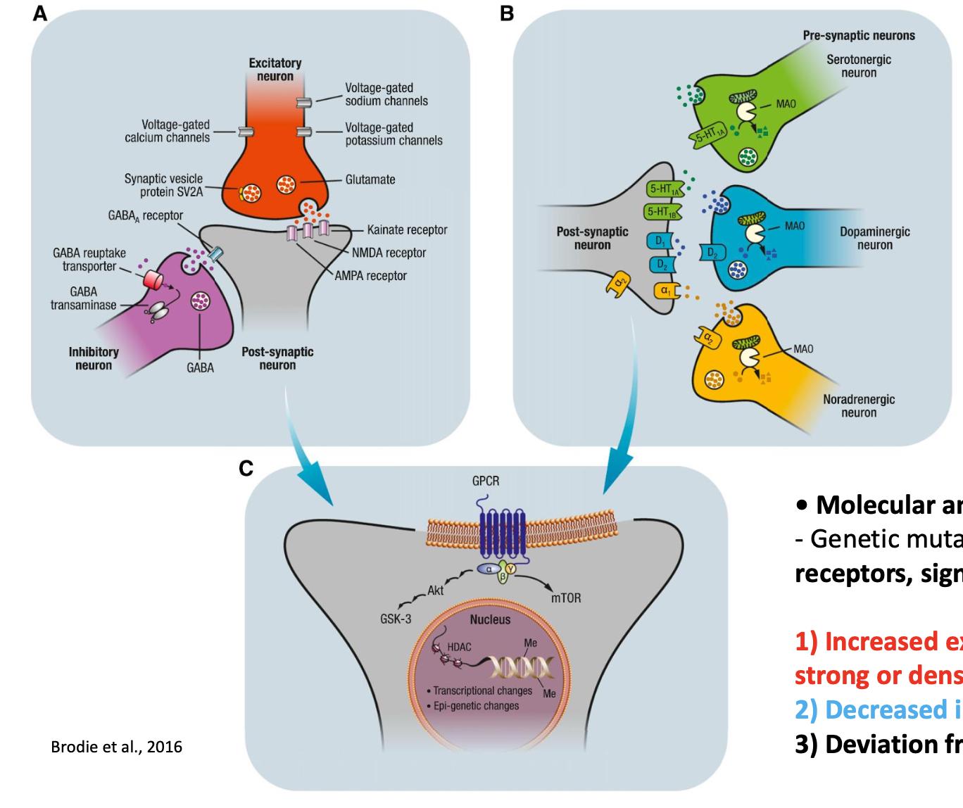
1) Increased excitation: mutations in sodium channels, excessively strong or dense excitatory connections. 2) Decreased inhibition: impairments in GABAergic synaptic inhibition 3) Deviation from delicate excitation-inhibition balance (E-I balance)
2. Sleep
- Sleep
- Readily reversible state of reduced responsiveness to the environment
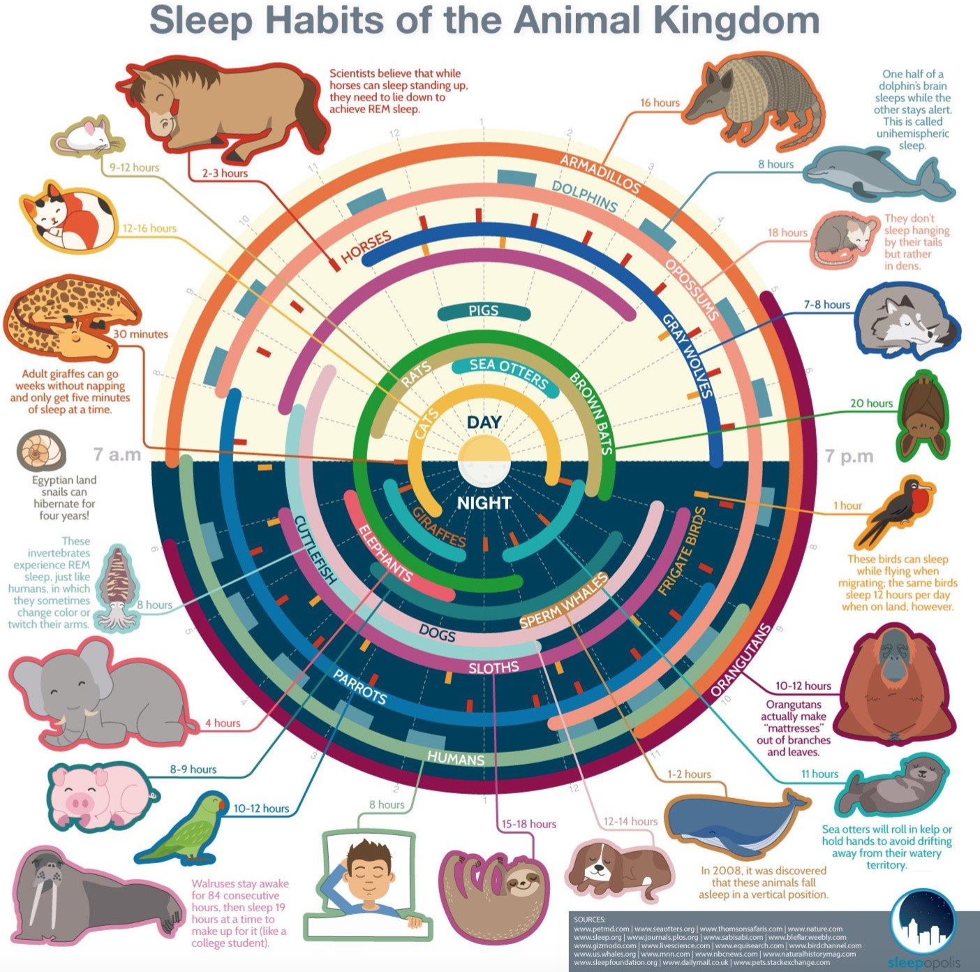 (https://infographicjournal.com/sleep-habits-of-the-animal-kingdom/)
Why do we sleep? - Still inconclusive.
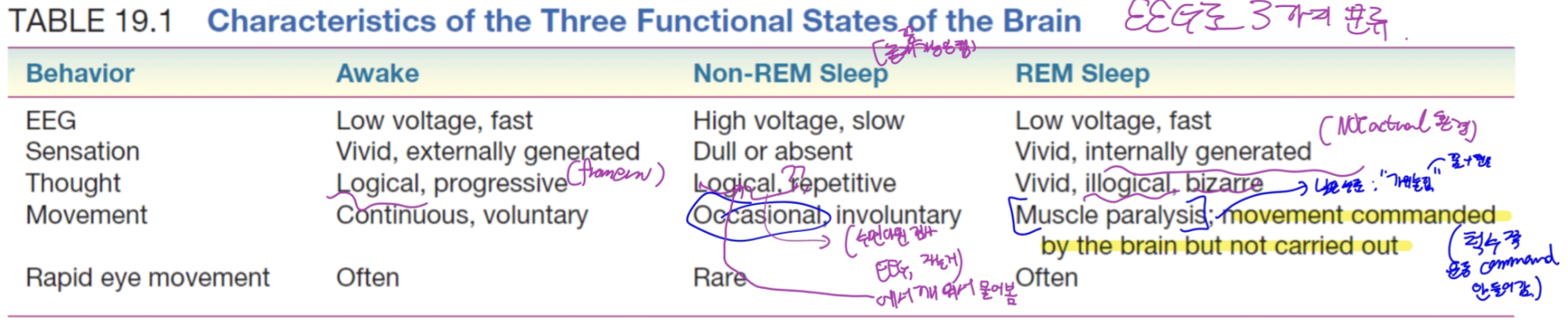
2.1. Three Functional States of the Brain
Dement’s saying
1) Awake 2) Non-REM Sleep
An idling brain in a movable body.
- The lowest level of brain functions (energy use, firing rates, mental processes)
- Activation of parasympathetic division of the ANS.
- No recall of vivid dreams.
- Slow, large amplitude of EEG rhythms => high synchrony of cortex.
- Most sensory input cannot reach the cortex.
3) REM Sleep
An active, hallucinating brain in a paralyzed body
- recall of detailed episodes of dreams
- Activation of sympathetic division
- Fast, low voltage fluctuations (like waking brain)
- atonia; an almost total loss of skeletal muscle tone. (muscle paralysis)
- Except: muscle controlling eye movement and the tiny muscles of the inner ear.
- Body’s temperature control system quits (inexplicable)
근육긴장도(muscle tone)는 수동적인 신장에 대한 근육의 저항으로 정의된다.
movie: REM sleep
2.2. The Sleep Cycle
Cycle of non-REM and REM sleep: every 90 munutes, with REM sleep getting more and more longer.
Best measure of successful sleep is the quality of your time awake.
In my case, I’d say an 8 hours of sleep is the best sleep time.
2.3. Why Do We Sleep?
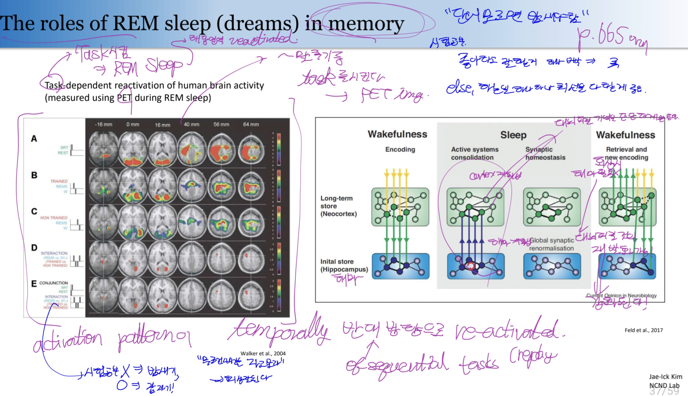
2.4. Functions of dreaming & REM Sleep
- REM sleep
- REM rebound: trying to maintain REM sleep.
- REM deprivation: No major psychological harm during the daytime.
REM sleep somehow aids the integration or consolidation of memories.
cf.
- Depriving humans or rats of REM sleep: impair their ability to learn a variety of tasks.
-
After intense learning, REM sleep duration Increase
- Dreaming
- Disguised wish fulfillment (Sigmund Freud): unconscious way to express our sexual and aggressive fantasies.
- Activation-synthesis hypothesis
- : Dreams are seen as the associations and memories of the cerebral cortex elicited by the random discharges of the **pons** during REM sleep.
- Hence the pontine neurons (via thalamus) activate various area of the cerebral cortex, eliciting images and emotions.
- As the cortex tries to synthesize the disparate images into a sensible whole, It does predict the bizarre and nonsensical dreams.
- Drawback: Cannot explain how random activity can trigger the complex and fluid stories that may dreams contain.
2.5. Neural Mechanisms of Sleep
- Localizing; Finding the area that triggers sleeping
- Lesion: removed => function changes.
- Stimulation: Changes after activations.
- Recording: Activity-brain state relationship.
.png)